


Many of the greatest discoveries in history were not planned—they happened by accident. One of the most powerful examples is the discovery of X-rays in 1895 by the German physicist Wilhelm Conrad Röntgen.
While experimenting with cathode rays in his laboratory, Röntgen noticed a strange glow coming from a fluorescent screen nearby. This unexpected observation would soon lead to one of humanity’s most transformative scientific breakthroughs: X-rays. For the first time in history, doctors could see inside the human body without performing surgery. This moment marked the birth of medical imaging, forever changing the practice of medicine.
Within just a few months, hospitals around the world began using X-rays to diagnose broken bones, detect tumors, and identify internal diseases with unprecedented accuracy. What started as a curious laboratory anomaly quickly became an essential medical tool.
The impact of X-rays extended far beyond medicine. They revolutionized physics, engineering, and security, deepening our understanding of energy, matter, and atomic structure. X-ray research laid the groundwork for nuclear physics and modern electronics. Röntgen’s unexpected discovery opened a window into the invisible world, reminding us that curiosity and careful observation can change the course of history.
This article explores how X-rays were discovered, the life of Wilhelm Conrad Röntgen, the scientific background that shaped his work, the accidental circumstances behind the discovery, and the far-reaching consequences that followed. It also traces how X-ray technology evolved, the challenges it introduced, and the ways it continues to shape the modern world.
Table of Contents
Quick Facts
| Fact | Detail |
| Discovery Name | X‑Rays (Roentgen rays) |
| Discovered By | Wilhelm Conrad Röntgen (German physicist) |
| Discovery Date | November 8, 1895 |
| Location | Würzburg, Germany (University lab) |
| Experiment Context | Cathode ray experiments with Crookes tube |
| Initial Observation | Fluorescent glow on screen despite opaque shielding |
| First Famous Image | Hand X‑ray of Röntgen’s wife showing bones & wedding ring |
| Original Name | “X‑rays” — X for unknown |
| First Scientific Paper | Über eine neue Art von Strahlen (On a New Kind of Rays), Dec 1895 |
| Immediate Impact | Revolutionized medical diagnosis & imaging |
Wilhelm Conrad Röntgen: The Man Behind the Discovery
The story of X-rays cannot be separated from the life and character of Wilhelm Conrad Röntgen, whose curiosity and perseverance led to one of the greatest scientific discoveries in history.
Röntgen was born on March 27, 1845, in the town of Lennep, Germany (now part of Remscheid). He grew up in a modest household. His father was a cloth merchant, while his mother came from a well-educated Dutch family. From an early age, Röntgen showed a quiet but determined interest in understanding how things worked. Although he was not considered an outstanding student, he became known for his persistence, focus, and practical intelligence—qualities that would later define his success as an experimental physicist.
His educational path was not without obstacles. While studying at a technical school in Utrecht, Röntgen was unfairly accused of drawing a caricature of a teacher and was temporarily expelled. For a less determined person, this setback might have ended an academic career altogether.
Instead, Röntgen pressed on. He eventually gained admission to the Polytechnic Institute in Zurich, where he studied mechanical engineering. In 1869, under the guidance of the renowned physicist August Kundt, he earned his doctoral degree. During the early years of his career, Röntgen assisted Kundt in experiments involving air, light, and electricity. These experiences sharpened his laboratory skills and instilled in him a deep respect for precision and careful measurement.
Over the next two decades, Röntgen held academic positions at several universities, including Strasbourg, Giessen, and finally Würzburg, where he would make his historic discovery. Colleagues often described him as a man of few words—methodical, humble, and deeply committed to his research. He preferred working alone in his laboratory, often late into the night, driven not by fame or recognition but by pure scientific curiosity.
By the early 1890s, Röntgen was experimenting with cathode rays and Crookes tubes, a field that fascinated many physicists at the time. His goal was to understand how these invisible electrical emissions behaved under different conditions. What he did not realize was that these seemingly routine experiments were about to reveal an entirely new form of radiation—one capable of passing through solid objects and exposing the hidden structures within.
In many ways, Röntgen’s life represents the perfect blend of discipline, curiosity, and openness to the unexpected. His patience and attention to detail created the conditions for a discovery that would permanently transform science and medicine. What began as a quiet observation in a darkened laboratory became a turning point in human history.
The Scientific World Before the Discovery

To fully understand the importance of Wilhelm Conrad Röntgen’s discovery, we must first look at the scientific landscape of the late nineteenth century. This was a period marked by intense curiosity and rapid progress in the study of electricity, magnetism, and light. Across Europe, laboratories buzzed with experiments that sought to uncover the hidden forces shaping the physical world.
Scientists of the time were particularly fascinated by mysterious invisible phenomena they broadly referred to as “rays” or “radiation.” Although they did not yet realize it, these investigations were laying the foundations of modern physics and atomic theory. Researchers were slowly discovering that reality extended far beyond what the human eye could see.
One of the most important experimental tools of this era was the Crookes tube, named after the British scientist Sir William Crookes. This glass tube, from which most of the air had been removed to create a partial vacuum, contained two metal electrodes connected to a high-voltage power source. When electricity passed through the tube, it produced strange glowing effects. These emissions became known as cathode rays, and their unusual behavior captivated scientists. The rays appeared to travel in straight lines, cause certain materials to glow, and interact with matter in unexpected ways.
Prominent physicists such as Heinrich Hertz, Philipp Lenard, and J. J. Thomson devoted significant effort to studying these rays. Hertz demonstrated that cathode rays could pass through thin metal foils, while Lenard designed tubes with thin aluminum windows that allowed the rays to exit the glass so their behavior could be studied outside the tube. These findings suggested that some form of invisible radiation could penetrate solid materials, challenging traditional ideas about matter and light. Yet the true nature of these rays remained unclear.
By this time, James Clerk Maxwell had already established the theory of electromagnetic radiation. However, scientists had not yet fully grasped that radiation extended beyond visible light into a vast spectrum that included ultraviolet rays, X-rays, and gamma rays. Experimental equipment was still relatively crude, and researchers often relied more on careful observation than on fully developed theoretical frameworks.
It was within this environment—defined by rigorous experimentation, limited understanding, and constant surprise—that Röntgen’s work emerged. Like many of his contemporaries, he was experimenting with cathode rays in vacuum tubes. Unlike most others, however, Röntgen paid extraordinary attention to anomalies. He tested every irregularity instead of dismissing it. This habit allowed him to notice something astonishing: a new type of radiation capable of passing through wood, books, and even human flesh.
The scientific groundwork had been laid. The world stood on the edge of a major discovery—not one deliberately engineered, but one waiting to be noticed in the quiet of a laboratory in Würzburg.
The Accidental Discovery of X-Rays
On the evening of November 8, 1895, Wilhelm Conrad Röntgen was working alone in his modest laboratory at the University of Würzburg in Germany. The air was cold and still, broken only by the faint hum of electrical equipment. Röntgen was experimenting with a Crookes tube—a partially evacuated glass tube designed to study cathode rays. His objective that night was simple: to observe how these rays behaved when surrounded by different materials.
To eliminate any interference from visible light, Röntgen had carefully wrapped the tube in black cardboard, ensuring complete darkness. As he passed a high-voltage electric current through the tube, something unexpected happened. A few feet away, on a nearby bench, he noticed a faint, shimmering glow. The source was a small screen coated with barium platinocyanide, a substance known to fluoresce when exposed to certain types of radiation.
The glow made no sense. The screen was too far away to be affected by ordinary light or cathode rays, which could not pass through the cardboard covering the tube. Something unseen was traveling through the air and striking the screen. Intrigued, Röntgen began a series of systematic experiments. He placed various objects—wood, books, and metal plates—between the tube and the screen. The mysterious glow persisted, though its intensity varied depending on the material.
Then came the moment that would change history. When Röntgen placed his own hand between the tube and the screen, he saw the shadowy outline of his bones projected onto the glowing surface. His flesh appeared almost transparent, while his bones stood out dark and sharp. He had discovered a new, invisible, and extraordinarily powerful form of radiation—one that could penetrate materials opaque to ordinary light.
Over the following weeks, Röntgen worked in near-total secrecy. Before announcing his findings, he wanted absolute certainty. He repeated his experiments meticulously, studying how the new rays behaved when passing through metals, glass, paper, and even water. He observed that denser materials absorbed more of the radiation, while lighter materials allowed it to pass through more easily.
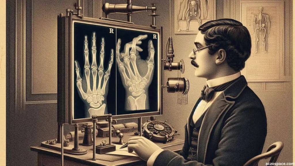
To document his results, Röntgen used photographic plates to capture permanent images of the shadows produced by the rays. On December 22, 1895, he created what would become one of the most famous scientific images in history: an X-ray photograph of his wife Anna Bertha’s hand. The image clearly revealed her bones and the dark outline of her wedding ring. Upon seeing it, she reportedly exclaimed, “I have seen my death!” This haunting photograph marked the first X-ray image of a human body part and symbolized the dawn of a new era in science and medicine.
Röntgen referred to the mysterious radiation as “X-rays,” using the letter X to signify the unknown. He soon prepared a detailed scientific paper titled “On a New Kind of Rays” (Über eine neue Art von Strahlen), which he presented to the Würzburg Physical-Medical Society in late December 1895. The paper was published in January 1896, accompanied by the astonishing images that left the scientific community stunned.
Within days, news of the discovery spread across Europe and the United States, provoking equal parts excitement and skepticism. Scientists rushed to replicate Röntgen’s experiments, while newspapers around the world celebrated the revelation of a technology that seemed almost magical.
The discovery of X-rays was not mere luck. It was the result of Röntgen’s discipline, patience, and relentless curiosity. Where others might have dismissed the faint glow as a laboratory oddity, Röntgen investigated it with care and persistence. His approach reflected the true spirit of science.
That single evening in November 1895 marked a turning point in human history. With the discovery of X-rays, the invisible became visible. For the first time, humanity could peer inside the human body and beyond, revealing hidden structures that had remained unseen for millennia.
Early Documentation and the First Experiments
By the end of 1895, news of Wilhelm Conrad Röntgen’s astonishing discovery sent waves of excitement, confusion, and amazement through the scientific world. An invisible form of radiation that could pass through flesh and reveal the hidden structures of bones and metal objects was something no one had ever witnessed before. To describe this unknown phenomenon, Röntgen chose the mathematical symbol “X,” long used to represent the unknown, and named them X-rays. The term quickly gained global acceptance, although in countries such as Germany, they were also called “Röntgen rays” in his honor.
Within weeks of Röntgen’s announcement, laboratories around the world began reproducing his experiments. Scientists in France, Britain, the United States, and Russia successfully generated the same mysterious rays using their own Crookes tubes. By early 1896, scientific journals were filled with reports of new X-ray images—hands, coins, bullets lodged in limbs, and even small animals. It was clear that an entirely new branch of science had been born almost overnight.
Early Public Demonstrations
Röntgen himself remained cautious and deeply modest. Believing that scientific knowledge should belong to all humanity, he refused to patent his discovery. Others, however, were eager to showcase the new technology. In early 1896, physics professors across Europe began giving public lectures featuring live X-ray demonstrations. Crowds gathered to watch the eerie projection of their own bones glowing on fluorescent screens. Newspapers described the experience as nothing less than “seeing the invisible.”
In Paris, the physicist Henri Becquerel became fascinated by X-rays, and while studying related phenomena later that year, he accidentally discovered radioactivity. In London, scientists at the Royal Institution produced numerous X-ray images that captured the imagination of doctors and the general public alike. What had begun as a laboratory curiosity rapidly entered mainstream public awareness, transforming how people thought about the limits of human perception.
The First Medical Applications
Almost immediately, physicians recognized the enormous medical potential of Röntgen’s discovery. Before X-rays, diagnosing broken bones, swallowed objects, or internal tumors relied heavily on painful probing and educated guesswork. Now, a single image could reveal what lay beneath the skin—without a single incision.
In January 1896, just one month after Röntgen published his paper, a Scottish physician named John Macintyre carried out the first medical X-ray examination in the United Kingdom. He successfully located a coin that a patient had accidentally swallowed. Soon afterward, hospitals across Europe and North America began installing X-ray equipment. For wounded soldiers, particularly those injured in battle, doctors could now locate bullets and fractures before surgery. Pain was reduced, surgeries became more precise, and countless lives were saved. A medical revolution had begun almost overnight.
Fascination, Myths, and Misunderstandings
The public’s fascination with X-rays also gave rise to exaggeration and misinformation. Some people believed the rays could read minds or see through clothing. Entrepreneurs marketed so-called “X-ray-proof underwear,” while photographers in major cities opened X-ray studios offering curious customers images of their own skeletons. Newspapers published cartoons poking fun at the idea that privacy itself had become obsolete.
Although many of these claims were wildly exaggerated, they reflected how mysterious and unsettling the discovery seemed at the time. Very few people truly understood the nature of X-rays. Some believed they possessed magical or psychic powers. Even scientists were unsure whether X-rays were a form of light, a stream of particles, or something entirely new.
The Dawn of a New Era
Within months of Röntgen’s announcement, X-rays had become a powerful tool across multiple fields—including science, medicine, and even art. Early experimenters used them to study the internal structures of plants, insects, and metals. Artists were inspired by the haunting beauty of X-ray images, while engineers employed them to inspect machinery and detect hidden structural flaws invisible to the naked eye.
Yet behind the excitement lurked a danger that few initially recognized. Early X-ray operators were often exposed for long periods and suffered burns and injuries as a result. At the time, no one fully understood that radiation could damage living tissue. It would take years of painful experience before the medical risks of X-rays were acknowledged and proper safety precautions were introduced.
Still, in those first months of 1896, the world gazed in awe at Röntgen’s remarkable rays. They had opened a window into the unseen, transforming science, medicine, and human imagination forever—and redefining what it meant to truly see.
Scientific Reaction and Global Spread
News of Wilhelm Conrad Röntgen’s extraordinary X-rays spread faster than almost any scientific discovery of the nineteenth century. By early 1896—just two months after Röntgen published his paper—the entire scientific world was captivated. From Europe to North America and Asia, physicists, physicians, and engineers rushed to their laboratories, eager to reproduce the strange effects using cathode-ray tubes. Newspapers described the discovery as “a miracle of the modern age,” capturing the public’s sense of awe.
A Wave of Experiments Across the World
Scientific institutions and universities quickly moved to verify Röntgen’s findings. In Britain, Sir William Crookes, whose work on vacuum tubes had paved the way for the discovery, successfully confirmed the existence of X-rays and praised Röntgen’s careful, methodical approach. In France, Henri Poincaré and Henri Becquerel conducted their own experiments. By the end of that same year, Becquerel would go on to discover natural radioactivity, a breakthrough directly inspired by the study of X-rays.
Across the Atlantic, the famously inventive Thomas Edison turned his attention to the new rays. He began designing improved X-ray devices and experimenting with fluorescent materials to enhance image clarity. Edison’s involvement helped push the technology toward practical applications and wider public awareness.
One reason X-rays spread so rapidly was their relative simplicity. Almost any well-equipped physics laboratory could generate them using a Crookes tube, a Ruhmkorff coil to produce high voltage, and photographic plates. Within weeks, laboratories across Europe and America were producing X-ray images and sharing them through scientific journals and popular newspapers alike.
Scientific Amazement and Recognition
Röntgen’s meticulously written paper impressed even the most skeptical scientists. Titled “On a New Kind of Rays,” it became a model of scientific clarity and restraint. Most importantly, it presented photographic evidence that could not be easily dismissed. Images of bones, coins, and metal objects transformed disbelief into admiration.
Prestigious institutions such as the Royal Society in London and the French Academy of Sciences quickly recognized the importance of the discovery. Many leading physicists described X-rays as the greatest breakthrough since electricity itself. In a world just beginning to understand electromagnetism, the idea that invisible rays could pass through solid matter opened entirely new dimensions in physical science.
Röntgen’s Recognition and Humility
Despite the sudden fame, Röntgen remained deeply modest. He avoided publicity and refused to patent his discovery, believing that scientific knowledge should benefit all of humanity. Later in life, he even donated his Nobel Prize money to his university. When asked why he called the new radiation “X-rays,” his answer was characteristically simple: “Because I did not know what they were.”
In 1901, Röntgen was awarded the first Nobel Prize in Physics, honoring his “extraordinary services” in discovering the rays that would later bear his name. Even then, he downplayed his achievement, insisting that he had merely continued his experiments and that the discovery was the result of persistence combined with chance.
Rapid Expansion in Medicine and Industry
By the late 1890s, X-rays were already transforming medical practice worldwide. In Britain, France, and the United States, hospitals routinely used them to diagnose fractures, locate bullets, and detect internal conditions that had previously gone unseen. During wartime, mobile X-ray units were deployed close to battlefields, allowing surgeons to operate with far greater precision and saving countless lives.
Beyond medicine, scientists began using X-rays to study the internal structure of crystals and metals. This work led to the development of X-ray crystallography, a field that would later play a crucial role in revealing the structure of DNA and other complex molecules. Engineers used X-rays to detect hidden flaws in machinery and pipelines. Artists and archaeologists adopted the technology to examine paintings, fossils, and ancient artifacts without causing damage.
A Discovery That United the World
Röntgen’s discovery proved to be something rare in human history. It united scientists, doctors, and the general public in a shared sense of wonder. It demonstrated how careful observation—combined with curiosity and a touch of chance—could uncover entirely new worlds hidden within the ordinary. X-rays bridged the gap between physics and medicine, theory and application, becoming one of the earliest examples of truly interdisciplinary science.
In less than a year, X-rays traveled from a quiet laboratory in Würzburg to every major continent on Earth. For the first time, humanity could see the invisible—not through imagination or myth, but through the power of science itself.
Impact on Medicine and Modern Science
The discovery of X-rays in 1895 did far more than introduce a new scientific phenomenon. It fundamentally reshaped the relationship between science and human life. For the first time in history, doctors could look inside the human body without performing surgery. What began as a laboratory curiosity quickly became one of the most powerful diagnostic tools ever developed. Its influence on medicine, physics, chemistry, and biology has continued to shape modern science for more than a century.
A Revolution in Medical Diagnosis
Before Röntgen’s discovery, diagnosing internal injuries was often a matter of educated guesswork. Broken bones, swallowed objects, and internal bleeding could only be confirmed through painful, invasive procedures. Within months of the announcement of X-rays, hospitals across Europe and North America began using X-ray machines to capture images of bones and internal organs.
This new technology allowed doctors to:
- Clearly identify fractures
- Locate bullets or metal fragments inside the body.
- Detect hidden infections and tumors with far greater accuracy.
By 1896, just a year after Röntgen’s paper was published, medical journals in Britain and the United States were filled with X-ray images. Surgical procedures became more precise, safer, and less invasive, marking a turning point in patient care.
Dentistry was transformed as well. Dentists began using X-rays to detect cavities, root infections, and bone damage that were previously invisible. More than a century later, this early form of dental radiography remains a standard practice worldwide.
The Birth of Radiology
Röntgen’s discovery gave rise to an entirely new medical specialty: radiology. Physicians trained in interpreting X-ray images became known as radiologists, developing techniques to distinguish between healthy and diseased tissue. As the field grew, new equipment was designed to control exposure time, improve image clarity, and protect both patients and medical staff from excessive radiation.
By the early twentieth century, hospitals had established dedicated radiology departments. A new profession—radiologic technologists—emerged, specializing in operating imaging equipment. Scientists also developed contrast agents that made blood vessels and organs more visible under X-rays, allowing doctors to map the body with unprecedented detail.
Radiation Therapy and Cancer Treatment
Another unexpected outcome of Röntgen’s discovery was the use of radiation to treat disease. Shortly after X-rays were discovered, scientists such as Marie and Pierre Curie identified radioactive elements like radium and polonium. Researchers soon realized that radiation could do more than reveal hidden structures—it could also destroy diseased cells.
By the early 1900s, physicians were experimenting with controlled doses of radiation to treat skin conditions and cancer. These early efforts evolved into radiation therapy, now one of the most important treatments in modern oncology. Today, millions of cancer patients around the world receive life-saving radiation treatments, a medical legacy that can be traced directly back to Röntgen’s work.
Expanding the Frontiers of Science
Beyond medicine, X-rays opened entirely new fields of scientific research. In physics, they revealed the internal structure of matter. In 1912, Max von Laue demonstrated that X-rays could be diffracted by crystals, proving that they behaved like electromagnetic waves with extremely short wavelengths. This discovery led to the development of X-ray crystallography, a revolutionary method for studying atomic and molecular structures.
Thanks to crystallography, scientists later uncovered the double-helix structure of DNA in 1953, one of the greatest scientific achievements of all time. Chemists used X-rays to analyze materials and identify new elements. Engineers applied them to inspect welds, detect flaws in aircraft and machinery, and improve industrial safety and precision.
A Lasting Legacy
More than a century later, X-rays remain at the heart of modern science and medicine. They form the foundation of advanced imaging technologies such as CT scans, fluoroscopy, and even airport security scanners. Every time a patient undergoes a chest X-ray or a doctor examines a fracture, the same fundamental principles discovered by Röntgen in 1895 are at work.
Röntgen’s discovery changed not only how humans see their own bodies, but also how they understand the universe. Born from an accident, it demonstrated that curiosity, patience, and careful observation can lead to discoveries that transform the world. The invisible was made visible—and modern science was never the same again.
Hidden Dangers and the Evolution of Safety
Although Wilhelm Conrad Röntgen’s discovery of X-rays was celebrated worldwide, its potential dangers were not immediately understood. In the early excitement of seeing bones and hidden objects through solid matter, scientists and doctors paid little attention to the long-term effects of radiation exposure. It took years of observation, experience, and painful lessons for the medical community to fully recognize the risks associated with X-rays.
Early Cases of Radiation Injury
Within months of the widespread adoption of X-rays, troubling reports began to surface. Scientists and medical staff who worked closely with the new rays experienced burns, hair loss, and persistent skin injuries. Physicians who regularly performed X-ray examinations often developed reddened skin, chronic wounds, and tissue damage. In some cases, prolonged exposure later led to serious conditions, including cancer.
These early injuries were largely the result of inadequate safety practices. Operators frequently held photographic plates by hand or positioned patients extremely close to X-ray tubes, unaware that the radiation could penetrate human tissue. Protective shields, lead aprons, and safe operating distances were not yet standard practice. Early laboratories also lacked reliable methods to control or measure exposure levels.
Recognizing the Risks
By the early twentieth century, researchers began to understand that X-rays are a form of ionizing radiation, capable of altering atomic structures within living cells. Scientific work by figures such as Marie Curie and geneticist Hermann Joseph Muller highlighted the long-term biological effects of radiation exposure. Their findings made it clear that caution was essential in both medical and research settings.
As awareness grew, the scientific and medical communities began introducing safety measures. Lead aprons, gloves, and protective barriers became standard equipment. X-ray machines were redesigned to focus radiation only on targeted areas of the body. Exposure times were shortened, and maintaining distance from the radiation source became a fundamental safety principle.
The Development of Radiation Safety Standards
By the mid-twentieth century, formal radiation protection standards were firmly in place. Regulatory organizations such as the International Commission on Radiological Protection (ICRP) and national health authorities established guidelines to limit exposure for both patients and professionals. Monitoring devices, including dosimeters, allowed workers to track cumulative radiation doses over time.
Modern X-ray systems now include multiple built-in safety features, such as:
- Automatic shutoff mechanisms
- Adjustable beam intensity and precise targeting controls
- Digital imaging technology, which reduces exposure while improving image quality
Today, the risks of X-rays are well understood, and when used responsibly, their benefits far outweigh the dangers.
Lessons Learned
The early misuse of X-rays serves as a powerful reminder that scientific breakthroughs often come with unforeseen risks. Röntgen’s accidental discovery brought extraordinary benefits to humanity, but it also highlighted the importance of careful study, regulation, and ethical responsibility. Even the most promising technologies must be evaluated for their potential impact on human health and safety.
Despite these early challenges, X-rays remain one of the most valuable tools in medicine, science, and industry. They have transformed our ability to see the invisible, diagnose disease, and explore the world at atomic and molecular scales. Most importantly, they continue to evolve—becoming safer, more precise, and more effective with each generation of innovation.
The Modern Evolution of X-Ray Technology
Since Wilhelm Conrad Röntgen’s accidental discovery in 1895, X-ray technology has undergone a remarkable transformation. What began with fragile glass tubes producing faint images over long exposure times has evolved into fast, highly precise digital systems capable of delivering detailed results in seconds. Modern innovations have not only improved image quality but have also dramatically reduced radiation exposure, making X-rays safer and more effective than ever before.

Digital X-Ray Imaging
The shift from traditional photographic plates to digital X-ray detectors marked a turning point in medical imaging. Instead of relying on film, digital sensors capture X-ray data electronically, allowing images to appear instantly on computer screens. Clinicians can store, share, and analyze these images with ease.
This transition has led to several major advantages:
- Fewer repeat scans, reducing unnecessary exposure
- Lower overall radiation doses
- Greater diagnostic accuracy
- Enhanced image adjustments for radiologists
- Easy zooming and side-by-side comparisons of areas of interest
Digital X-rays are now widely used in dentistry, orthopedics, and general medicine, providing clear and detailed views of bones, teeth, and internal organs. Because digital systems are efficient and portable, X-ray imaging has become more accessible—even in small clinics and mobile healthcare units.
Computed Tomography (CT)
One of the most significant advances following Röntgen’s discovery is computed tomography (CT). Developed in the 1970s, CT scanning uses X-rays taken from multiple angles to create detailed three-dimensional images of the body. Unlike standard X-rays, which produce flat images, CT scans generate cross-sectional views that reveal internal structures with exceptional clarity.
CT technology has transformed medical diagnosis. It allows doctors to detect tumors, internal bleeding, and organ abnormalities with far greater precision. Today, CT scans are indispensable in emergency medicine, oncology, cardiology, and neurology, enabling faster diagnoses and more effective treatment planning.
Fluoroscopy and Real-Time Imaging
Another important development is fluoroscopy, which provides real-time moving images of the body using X-rays. Surgeons and cardiologists rely on fluoroscopy to guide catheters, implants, and other instruments during procedures, significantly reducing risk and improving accuracy.
Modern fluoroscopy systems incorporate low-dose protocols that maintain image quality while minimizing radiation exposure. This balance between precision and safety reflects how far X-ray technology has advanced since its early days.
AI-Assisted X-Ray Analysis
In recent years, artificial intelligence (AI) has begun to play a growing role in X-ray interpretation. Machine-learning algorithms can now detect fractures, infections, tumors, and other abnormalities with impressive accuracy—often assisting radiologists by highlighting areas of concern more quickly than traditional methods.
AI also helps optimize imaging settings, reduce unnecessary exposure, and streamline clinical workflows. In developing countries and remote regions, AI-powered mobile X-ray units are proving especially valuable, delivering high-quality diagnostic support where trained specialists may be scarce. This collaboration between automation and human expertise represents a new frontier in medical imaging.
Industrial, Security, and Scientific Applications
Beyond medicine, X-ray technology plays a vital role in industry, security, and scientific research. Engineers use X-rays to inspect welds, pipelines, and aircraft components for hidden defects. At airports and border checkpoints, security agencies scan luggage and cargo to detect prohibited items and potential threats.
In archaeology and art conservation, X-rays allow researchers to examine fossils, artifacts, and paintings without causing damage. Scientists continue to rely on X-ray crystallography and related techniques to uncover the structures of molecules, proteins, and DNA—advancing chemistry, biology, and medical science alike.
A Technology That Continues to Evolve
From the faint glow in Röntgen’s darkened laboratory to today’s sophisticated imaging systems, X-ray technology has evolved far beyond what its discoverer could have imagined. Modern advancements emphasize safety, precision, and versatility, transforming an early experiment with cathode rays into one of humanity’s most powerful diagnostic and investigative tools.
The story of X-rays is ultimately a testament to curiosity and innovation. It shows how a single discovery can grow into a transformative technology—reshaping medicine, science, and industry, and continuing to expand our ability to see and understand the invisible world around us.
Conclusion
Wilhelm Conrad Röntgen’s accidental discovery of X-rays in 1895 remains one of the most remarkable moments in the history of science. What began as a modest laboratory experiment with a Crookes tube revealed an entirely new form of invisible radiation—one capable of passing through solid objects. That single discovery permanently reshaped medicine, physics, and modern technology.
X-rays revolutionized medical diagnosis almost overnight. For the first time, physicians could identify broken bones, locate foreign objects, and diagnose internal conditions without resorting to invasive procedures. This breakthrough laid the foundation for radiology, radiation therapy, and advanced imaging techniques such as CT scans and fluoroscopy. Beyond medicine, X-rays opened new pathways in physics, crystallography, and industrial inspection, influencing both scientific understanding and practical applications across the globe.
The journey from Röntgen’s faintly glowing screen to today’s AI-assisted imaging systems highlights the power of curiosity, careful observation, and persistence. It also serves as a reminder that every scientific breakthrough carries both opportunity and responsibility. Early radiation injuries led to the development of strict safety standards, ensuring that X-rays could be used effectively while protecting patients and professionals alike.
More than a century later, X-rays remain essential in medicine, science, industry, and security. Röntgen’s unexpected discovery continues to touch countless lives, proving that human knowledge can expand in profound ways through chance, innovation, and thoughtful refinement. It is a story of curiosity made visible—a spark that illuminated the unseen and fundamentally changed how humanity understands the world.
FAQs
Who discovered X-rays?
Wilhelm Conrad Roentgen discovered X-rays in 1895 during an experiment with cathode rays.
How was X-ray discovered by accident?
Roentgen noticed a fluorescent glow on a screen while testing tubes, revealing invisible rays that could pass through objects.