

The Untold X-Ray Discovery Story: How Wilhelm Röntgen Accidentally Changed the World Forever
Imagine discovering something invisible, something that can pass through solid objects and reveal what’s hidden beneath. That’s exactly what happened in one of the most surprising moments in scientific history. The X–ray discovery story begins not with a planned breakthrough, but with curiosity, experimentation, and a bit of accident.
In 1895, Wilhelm Conrad Röntgen stumbled upon a mysterious type of radiation while working in his laboratory. He didn’t set out to change medicine forever, but that’s exactly what he did. His discovery allowed humans, for the first time, to see inside the body without surgery, revolutionizing healthcare and opening new doors in science.
Table of Contents
In this article, you’ll explore the fascinating X–ray discovery story, learn how these invisible rays work, and discover their lasting impact on medicine, technology, and everyday life. Along the way, you’ll also uncover interesting facts that make this discovery truly unforgettable.
What Are X-Rays? A Simple Explanation
X-rays are a type of invisible energy, known as electromagnetic radiation, similar to visible light but with much higher energy. Because of this, they have the unique ability to pass through many materials that light cannot.
Here’s a simple way to understand how X-rays work:
- When X-rays are directed at an object, they pass through soft materials like skin and muscles.
- Denser materials, such as bones or metal, absorb more of the rays.
- This difference creates an image, allowing us to “see” inside objects or the human body.
That’s why, when you get an X-ray at a hospital, your bones appear clearly while softer tissues look lighter or less defined.
Everyday Uses of X-Rays
X-rays are not just used in hospitals; they are part of our daily lives in many ways:
- Medical Imaging: Doctors use X-rays to detect fractures, infections, and other conditions.
- Security Screening: Airports use X-ray scanners to check luggage for prohibited items.
- Industrial Inspection: Engineers use X-rays to examine machinery and structures without damaging them.
Even though they are invisible, X-rays play a powerful role in making modern life safer, healthier, and more advanced, all thanks to the incredible X-ray discovery story.
Who Was Wilhelm Röntgen?

Wilhelm Conrad Röntgen was a German physicist best known for discovering X-rays in 1895, one of the most important breakthroughs in scientific history. Born in 1845 in Germany, Röntgen grew up with a strong curiosity about how the natural world works, which later shaped his career in physics.
He studied mechanical engineering before moving into physics, eventually earning his PhD. Over time, he became a respected academic and researcher, known for his careful and precise experimental methods. His work often focused on understanding the behavior of energy and radiation, which ultimately led to his role in the famous X-ray discovery story.
Despite his groundbreaking achievement, Röntgen was a modest and private individual. He avoided publicity and refused to patent his discovery, believing that scientific knowledge should benefit all of humanity. His dedication, curiosity, and integrity continue to inspire scientists around the world.
His Scientific Journey Before the Discovery
Before the historic X-ray discovery story, Wilhelm Conrad Röntgen had already built a strong career in physics and education.
Work in Physics and Teaching Career
- Röntgen held professorships at several universities across Germany.
- He taught physics while conducting detailed experimental research.
- Known for his disciplined approach, he focused on accuracy and observation rather than speculation.
Research Focus Before X-Rays
- His earlier research explored electrical phenomena and the behavior of gases under low pressure.
- He conducted experiments with cathode rays, which were a major scientific topic at the time.
- These experiments laid the groundwork for his accidental discovery of X-rays.
Through years of dedication and curiosity-driven research, Röntgen unknowingly set the stage for one of the greatest discoveries in science, making his journey a crucial part of the X-ray discovery story.
The Accidental Discovery of X-Rays Explained
The most fascinating part of the X-ray discovery story is that it wasn’t planned; it happened completely by accident. In 1895, while experimenting in his laboratory, Wilhelm Conrad Röntgen made an observation that would change the world forever.
Here’s a simple step-by-step breakdown of that historic moment:
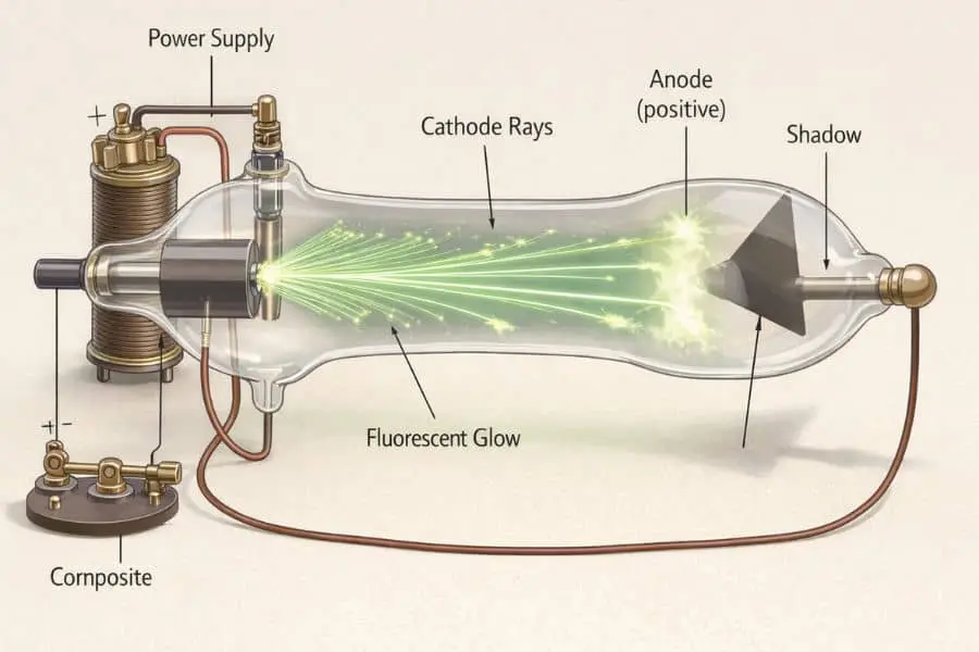
- Working with Cathode Rays
Röntgen was experimenting with a vacuum tube (called a Crookes tube), studying how cathode rays behaved in a dark room. - Covering the Tube
To block visible light, he covered the tube with black cardboard. This ensured that any visible glow wouldn’t interfere with his observations. - Unexpected Glow Appears
Even with the tube covered, he noticed a strange fluorescent glow coming from a nearby screen coated with a special chemical (barium platinocyanide). - Realization of Invisible Rays
This puzzled him; something invisible was traveling from the tube to the screen. These unknown rays could pass through the cardboard and air. - Testing Through Objects
Curious, Röntgen placed different objects between the tube and the screen. He discovered that the rays could pass through soft materials but were blocked by denser ones like metal and bone. - Naming the Discovery
Since he didn’t know what these rays were, he called them “X-rays,” with “X” representing something unknown.
This moment of curiosity and careful observation became the turning point in the X-ray discovery story, proving how unexpected discoveries can reshape the world.
The First X-Ray Image Ever Taken
One of the most iconic moments in the X-ray discovery story was the creation of the very first X-ray image.
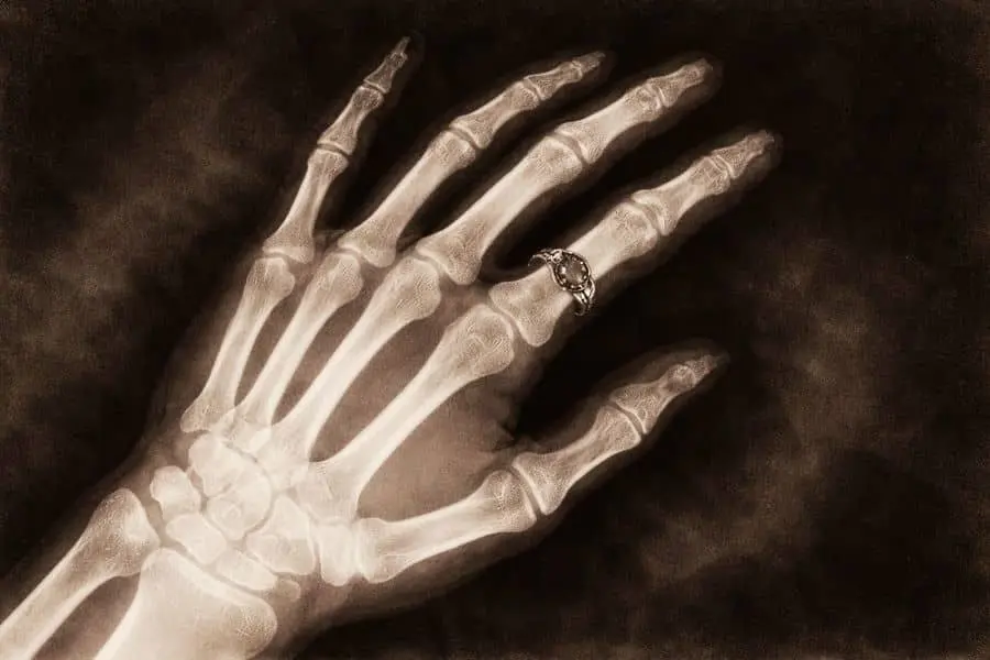
In his experiments, Wilhelm Conrad Röntgen asked his wife, Bertha Röntgen, to place her hand in front of the X-ray apparatus. The result was astonishing; a shadowy image showing the bones of her hand and her wedding ring clearly visible.
What Made This Image So Important?
- It was the first time in history that the inside of a human body was seen without surgery.
- The image proved that X-rays could be used for medical diagnosis.
- It marked the beginning of modern radiology.
Public Reaction
The reaction to this discovery was immediate and powerful:
- Scientists across the world were amazed and quickly began experimenting with X-rays.
- Doctors saw huge potential in diagnosing injuries without invasive procedures.
- The general public was both fascinated and slightly fearful of this mysterious new technology.
This single image turned the X-ray discovery story from a laboratory curiosity into a global scientific revolution, forever changing medicine and how we understand the human body.
Why the X-Ray Discovery Story Changed the World
The X-ray discovery story didn’t just introduce a new scientific concept; it transformed how humans understand and interact with the world. Almost immediately after Wilhelm Conrad Röntgen announced his discovery in 1895, its practical value became clear.
Before X-rays, doctors had to rely on physical examinations or perform surgery just to see what was happening inside the body. This was risky, painful, and often inaccurate. X-rays changed everything by allowing people to look inside the human body without making a single cut, marking the beginning of non-invasive diagnosis.
This breakthrough quickly spread across the globe, becoming one of the most important advancements in both science and medicine.
Impact on Medical Science

The medical field experienced the most immediate and life-changing benefits from the X-ray discovery story.
Detection of Fractures and Diseases
- Doctors could now clearly see broken bones without surgery.
- X-rays helped identify infections, tumors, and lung conditions.
- Early diagnosis became faster and more accurate.
Safer Diagnosis Compared to Surgery
- Reduced the need for exploratory surgeries.
- Lower risk for patients and faster recovery times.
- Improved treatment planning with precise internal images.
This innovation laid the foundation for modern diagnostic imaging techniques still used today.
Impact Beyond Medicine

While medicine benefited greatly, the X-ray discovery story also influenced many other industries.
Use in Engineering
- Engineers use X-rays to inspect the internal structure of machines.
- Helps detect cracks or weaknesses in materials without damaging them.
Industrial and Security Applications
- Airports use X-ray scanners to examine luggage and ensure safety.
- Factories rely on X-ray technology for quality control and product testing.
From hospitals to airports, the impact of X-rays continues to shape modern life, proving that this accidental discovery was truly world-changing.
Awards and Recognition
The groundbreaking contribution of Wilhelm Conrad Röntgen in the X-ray discovery story did not go unnoticed. In 1901, he was awarded the very first Nobel Prize in Physics by the Nobel Prize committee, an honor that recognized the immense impact of his discovery on science and humanity.
This achievement marked a historic moment, not just for Röntgen, but for the entire scientific community. His work opened the door to modern medical imaging and inspired countless advancements in physics and technology.
Global Recognition and Legacy
- Röntgen became internationally famous almost overnight after announcing X-rays.
- Universities and scientific institutions across the world honored his work.
- Despite his fame, he remained humble and avoided personal recognition.
- Today, his legacy lives on in hospitals, laboratories, and industries worldwide.
His discovery continues to save millions of lives, making him one of the most influential scientists in history.
Interesting Facts About the X-Ray Discovery Story
The X-ray discovery story is filled with fascinating details that make it even more remarkable:
- He Refused to Patent X-Rays
Wilhelm Conrad Röntgen chose not to patent his discovery, believing it should be freely available for the benefit of humanity. - Why the Name “X-Ray”?
He used the letter “X” to represent something unknown, as he didn’t fully understand the nature of the rays at the time. - Rapid Global Adoption
Within months of the discovery, scientists and doctors around the world began using X-rays for research and medical purposes. - First Nobel Prize Winner in Physics
Röntgen made history as the very first recipient of the Nobel Prize in Physics in 1901.
These facts highlight not only the scientific importance of the discovery but also the character and vision of the man behind it, making the X-ray discovery story truly unforgettable.
Are X-Rays Safe? (Modern Perspective)
X-rays are incredibly useful, but since they are a form of radiation, it’s natural to ask: are they safe? The answer is yes, when used properly and in controlled conditions. Modern science has made X-ray technology much safer than in the early days of the X-ray discovery story.
Understanding Radiation Risks
X-rays carry enough energy to affect atoms in the body, which means excessive exposure can potentially damage cells. However:
- The level of radiation used in most medical X-rays is very low.
- Occasional exposure (like getting an X-ray at a hospital) carries minimal risk.
- Problems typically arise only with frequent or high-dose exposure over time.
How Modern Technology Ensures Safety
Today’s medical systems are designed with safety as a top priority:
- Low-Dose Equipment: Modern machines use the smallest radiation dose necessary.
- Protective Shielding: Lead aprons and barriers protect sensitive parts of the body.
- Precise Targeting: X-rays are directed only at specific areas to limit exposure.
- Trained Professionals: Radiologists and technicians carefully control every procedure.
These advancements ensure that the benefits of X-rays far outweigh the risks in most situations.
Importance of Controlled Use
The key to safety lies in responsible and limited use:
- Doctors recommend X-rays only when medically necessary.
- Avoid unnecessary or repeated scans without proper reason.
- Always inform healthcare providers about previous imaging or pregnancy.
When used correctly, X-rays remain one of the safest and most powerful diagnostic tools in modern medicine; continuing the legacy of the X-ray discovery story in a responsible and life-saving way.
Conclusion
The X-ray discovery story is more than just a moment in scientific history; it’s a powerful reminder of how one unexpected observation can change the world forever. What began as a simple experiment by Wilhelm Conrad Röntgen in 1895 quickly became a breakthrough that transformed medicine, technology, and our understanding of the human body.
This discovery highlights the true power of curiosity. Röntgen didn’t ignore the strange glow he saw; he investigated it. That curiosity, combined with careful thinking and persistence, led to one of the greatest scientific achievements of all time.
Today, X-rays are part of our everyday lives, from hospitals and airports to industries and research labs. The next time you see an X-ray image or pass through a security scanner, it’s worth remembering the incredible journey behind it.
In the end, the X-ray discovery story teaches us an important lesson: sometimes, the biggest discoveries come from the simplest questions.
FAQs
Who discovered X-rays?
X-rays were discovered by Wilhelm Conrad Röntgen in 1895. While experimenting with cathode rays, he accidentally noticed invisible rays that could pass through solid objects, leading to the first X-ray image.
Why are X-rays called “X-rays”?
Röntgen named them “X-rays” because “X” represents the unknown. At the time of discovery, he didn’t understand the exact nature of these invisible rays, so the name reflected their mysterious qualities.